
|
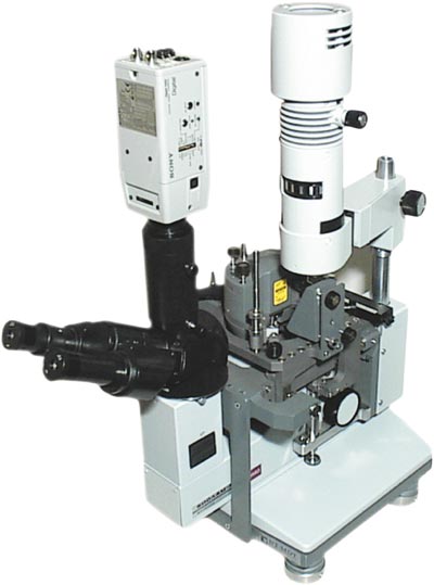
Solver P47BIO scanning probe microscope is a special device for biological and medical applications. It is based on a combination of an inverted optical and a scanning probe microscopes that enables visualisation of a sample surface by optical system during the scanning process in different SPM modes. An inverted optical microscope simplifies adjustment and positioning processes. The device provides investigations in ambient and liquid environment.
Measuring modes:
P47BIO applications include imaging of:
|  |
| Technical specification | |
| Sample size | 90x90 mm (max) |
| Scanning area | 60x60x3 µm, 100x100x5 µm. |
| Min scanning step | 0.06 A°, 0.1 A° |
| XY sample positioning | 30x30 mm (within 30 mm circle), 1 mm scale count. |
| Fine cantilever positioning | 4x4 mm range, 5 µm resolution. |
| Optical system | inverted microscope and color CCD camera SONY SSC-DC500P. |
| Tip viewing | scanning cantilever can be viewed on the monitor screen during the scanning procedure |
| Magnification | |
| with 14X eyepieces | 8.4X to 100X |
| with color CCD | 43X to 470X |
| Optical microscope: | |
| Resolution | 5 µm (with optional objective up to 1.5µm) |
| Field of view 14X eyepieces | 2.4 mm to 28mm |
| Scanning resolution | DAC down X, Y, Z and 16-bit ADC |
| Frequency range | 10 Hz-2 MHz |
| Accuracy of modulation frequency setting |
digital 32-bit (0.01 Hz). |
| Sample holder | |
| Vacuum chuck for Petri dishes | 60x60 mm and 90x90 mm |
| Holder with spring clamp for samples |
up to 30x100 mm |
| Inverted Optical Microscope | |
| Objectives | 2.5x0.08; 6.3x0.20; 10x0.22; 20x0.45 |
| Field of view | 0.5 mm to 6.2 mm |
| Magnification with eyepieces | 30X to 360X |
| Bright field, phase contract and polarization cotrast | |
| Trinocular head for CCD camera | |
|